 What is an ultrasound in pregnancy? A prenatal ultrasound (or sonogram) is a test during pregnancy that checks on the health and development of your baby. An obstetrician, nurse midwife or ultrasound technician (sonographer) performs ultrasounds during pregnancy for many reasons.
/mid-adult-female-doctor-using-ultrasound-scanner-1189207563-1d388c8128ad498c9f27407d4dc43eb6.jpg) Read on for a breakdown of the most common types of pregnancy ultrasounds, when you might get them, and what to expect during the prenatal scans.
Read on for a breakdown of the most common types of pregnancy ultrasounds, when you might get them, and what to expect during the prenatal scans.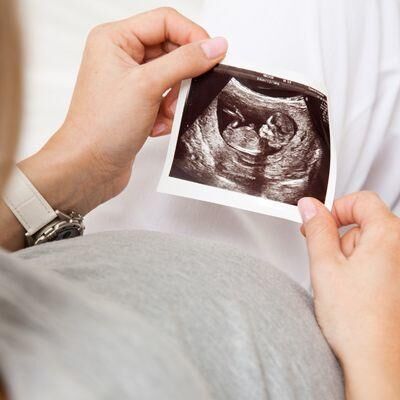 Here, learn what happens during an ultrasound in pregnancy, how many you might get and when, and why they’re so important for prenatal care. What is a pregnancy ultrasound? What is the purpose of a pregnancy ultrasound? How many ultrasounds during pregnancy will I get? Are pregnancy ultrasounds safe? How much do pregnancy ultrasounds cost?
 Vaginal ultrasound is done by inserting a wand-shaped transducer into your vagina. This type of ultrasound can provide better images in early pregnancy, enabling your provider to diagnose potential problems such as a miscarriage, a molar pregnancy, or an ectopic pregnancy.
 Early in pregnancy, ultrasounds are used to confirm the fetal heartbeat and the baby’s position in your uterus. Later, ultrasounds screen for fetal growth and placenta location, as well as baby's general health and anatomy.
 What is an Ultrasound Exam? This procedure uses high-frequency sound waves to scan a woman’s abdomen and pelvic cavity, creating a picture (sonogram) of the baby and placenta. Although the terms ultrasound and sonogram are technically different, they are used interchangeably and refer to the same exam.
 A pregnancy ultrasound is an imaging test that uses sound waves to create a picture of how a baby is developing in the womb (uterus). It is also used to check the female pelvic organs during pregnancy.